
Questions
TOX001 - Pediatric Gastric Decontamination Technique
Scenario
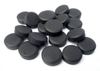
A 4-year-old child weighing 16 kg is brought to the pediatric emergency department 45 minutes following an unsupervised ingestion of an unknown quantity of his grandmother's carbamazepine tablets. The nursing officer prepares a slurry for enteral administration with the tablets depicted in the clinical photograph.
Questions
- Describe the primary biophysical mechanism of decontamination for the agent shown.
- List four absolute or relative contraindications to the administration of this decontamination therapy.
- Enumerate three documented clinical complications associated with its enteral delivery.
- Identify four xenobiotics for which multiple-dose administration of this agent (MDAC) significantly enhances systemic clearance, and outline the initial and subsequent pediatric dosing regimens.
Answer
- Mechanism of Action:
- Adsorption of dissolved xenobiotics within the gastrointestinal lumen across a large internal surface area ($950\text{--}2000\text{ m}^2/\text{g}$) via non-specific, reversible van der Waals forces, hydrogen bonding, and electrostatic interactions.
- Prevents systemic enteral absorption and interrupts enterohepatic or enteroenteric recirculation by establishing a downward concentration gradient from mesenteric capillaries into the gut lumen ("gut dialysis").
- Contraindications:
- Depressed conscious level or loss of protective airway reflexes without prior definitive endotracheal intubation.
- Ingestion of caustic, corrosive substances (strong acids or alkalis) due to risk of emesis, obscurement of mucosal visualization on endoscopy, and increased perforation hazard.
- Ingestion of pure hydrocarbons (risk of lethal chemical pneumonitis if aspirated).
- Gastrointestinal tract perforation, ileus, or mechanical bowel obstruction.
- Ingestants poorly adsorbed by activated charcoal: heavy metals (iron, lead, lithium), elemental electrolytes, alcohols, and ethylene glycol.
- Clinical Complications:
- Pulmonary aspiration pneumonitis and fatal bronchospasm or asphyxiation.
- Mechanical gastrointestinal obstruction, small bowel bezoar formation, or secondary perforation.
- Severe dehydration, hypernatremia, or osmotic shifts (when compounded with repeated doses of sorbitol-containing cathartics).
- Multiple-Dose Activated Charcoal (MDAC) Protocol:
- Target Xenobiotics (mnemonic: "These People Can't Drink"):
- Phenobarbital
- Theophylline
- Carbamazepine
- Dapsone
- Quinine
- Pediatric Dosing:
- Initial dose: $1\text{ g/kg}$ orally or via nasogastric tube (maximum $50\text{ g}$).
- Subsequent doses: $0.25\text{--}0.5\text{ g/kg}$ every $2\text{--}4\text{ hours}$ (maximum $25\text{ g/dose}$), titrated to bowel sounds and clinical status, discontinuing when clinical toxicity abates and cathartics avoided in repeat cycles.
- Target Xenobiotics (mnemonic: "These People Can't Drink"):
TOX002 - Systematic Selection of Specific Antidotes
Scenario
A clinical toxicologist is reviewing antidote stock requirements and treatment algorithms for the pediatric intensive care unit. You are asked to establish evidence-based guidelines regarding specific antidotes and their clinical indications.
Questions
- Enlist two definitive xenobiotic exposures or toxicological scenarios where Glucagon serves as an antidote, stating its molecular mechanism.
- Outline three separate toxicological indications for Pyridoxine (Vitamin $B_6$) therapy.
- List two non-acetaminophen toxic exposures where N-Acetylcysteine (NAC) provides therapeutic utility.
- Compare Calcium Disodium Edetate ($\text{CaNa}_2\text{EDTA}$) and Dimercaptosuccinic acid (DMSA / Succimer) in terms of route of administration, primary indications, and central nervous system safety in lead encephalopathy.
Answer
- Glucagon Indications and Mechanism:
- Indications:
- Severe $\beta$-adrenergic antagonist (beta-blocker) overdose refractory to crystalloids and vasopressors.
- Severe calcium channel blocker (CCB) toxicity with refractory bradycardia and cardiogenic shock.
- Severe sulfonylurea-induced refractory hypoglycemia (adjunctive temporary rescue).
- Mechanism:
- Bypasses blocked $\beta$-adrenergic receptors by activating independent myocardial glucagon-specific G-protein-coupled receptors, directly stimulating adenylate cyclase to increase intracellular cyclic AMP (cAMP), thereby augmenting chronotropy and inotropy.
- Indications:
- Indications for Pyridoxine (Vitamin $B_6$):
- Acute Isoniazid (INH) overdose presenting with status epilepticus and metabolic acidosis.
- Monomethylhydrazine-containing mushroom toxicity (Gyromitra esculenta false morel ingestion).
- Ethylene glycol toxicity (acts as an essential cofactor to divert glyoxylic acid conversion towards the benign metabolite glycine).
- Non-Acetaminophen Indications for N-Acetylcysteine:
- Carbon tetrachloride ($\text{CCl}_4$) and chloroform-induced acute hepatotoxicity (scavenges toxic trichloromethyl free radicals and replenishes hepatic glutathione).
- Inhalation injury from phosgene, chlorine gas, or blister agents (sulfur mustard) causing oxidative pulmonary epithelial injury.
- Non-acetaminophen-induced pediatric acute liver failure (idiopathic or viral) for improving systemic microcirculatory perfusion and oxygen extraction.
- Comparison of $\text{CaNa}_2\text{EDTA}$ vs Succimer (DMSA):
- Route of Administration:
- $\text{CaNa}_2\text{EDTA}$: Parenteral only (intravenous infusion or deep intramuscular with procaine).
- DMSA (Succimer): Enteral (oral capsules).
- Primary Clinical Indications:
- $\text{CaNa}_2\text{EDTA}$: Severe acute lead toxicity (blood lead level $\ge 70\ \mu\text{g/dL}$ or lead encephalopathy); zinc and manganese toxicity.
- DMSA: Moderate-to-severe lead poisoning without encephalopathy (blood lead level $45\text{--}69\ \mu\text{g/dL}$); acute arsenic and inorganic mercury toxicity.
- CNS Safety in Encephalopathy:
- $\text{CaNa}_2\text{EDTA}$ can mobilize lead from skeletal stores into extracellular fluid; if given as monotherapy in high lead burdens or encephalopathy, it may precipitate acute cerebral edema by redistributing lead across the blood-brain barrier. Hence, it must ALWAYS be preceded by Dimercaprol (British Anti-Lewisite / BAL) by at least 4 hours.
- DMSA is hydrophilic, does not mobilize bone lead into brain parenchyma, but cannot be used enterally in active encephalopathy with absent airway protection or vomiting.
- Route of Administration:
TOX003 - Domestic Enclosed Space Unconsciousness
Scenario
A 6-year-old child and both parents are brought to the emergency department during winter after being found stuporous in their native bedroom where an unserviced internal combustion generator was operating in an adjoining unventilated utility room. On examination, the child is lethargic, displays peripheral cutaneous flushing, pulse rate is $138/\text{min}$, respiratory rate is $28/\text{min}$, and pulse oximeter shows $\text{SpO}_2\ 99\%$ on ambient room air.
Questions
- Explain the physiological discrepancy between the pulse oximeter saturation reading ($\text{SpO}_2$) and actual arterial oxygen content ($C_aO_2$) in this clinical condition.
- Give the expected elimination half-life ($t_{1/2}$) of the toxic adduct under:
- Ambient room air ($21\%\ \text{FiO}_2$).
- $100\%$ normobaric oxygen delivered via high-flow non-rebreather reservoir mask.
- Hyperbaric oxygen therapy (HBOT at $2.5\text{--}3.0\text{ atmospheres absolute [ATA]}$).
- Identify the classic neuroimaging abnormality observed on magnetic resonance imaging (MRI) of the brain in severe toxicity.
- State three absolute indications for transferring a pediatric patient for Hyperbaric Oxygen (HBOT) therapy.
Answer
- Physiological Basis of Normal $\text{SpO}_2$ with Reduced Arterial Oxygen Content:
- Standard dual-wavelength pulse oximeters measure light absorption at $660\text{ nm}$ (red) and $940\text{ nm}$ (infrared).
- Carboxyhemoglobin (COHb) exhibits an optical absorption spectrum at $660\text{ nm}$ virtually identical to oxyhemoglobin ($\text{HbO}_2$). The pulse oximeter erroneously interprets COHb as $\text{HbO}_2$, producing a falsely normal or high $\text{SpO}_2$ reading.
- Actual fractional oxyhemoglobin saturation ($\text{SaO}_2$) and dissolved oxygen content are depressed. Carbon monoxide binds to hemoglobin with an affinity 200–250 times that of oxygen, shifting the oxyhemoglobin dissociation curve to the left (Haldane effect), which severely impairs peripheral oxygen unloading to tissues.
- Elimination Half-Life ($t_{1/2}$) Kinetics: $$\begin{aligned} t_{1/2}\text{ on Room Air } (21\%\ \text{FiO}_2) &= \mathbf{240\text{--}320\text{ minutes}} \quad (4\text{--}5.5\text{ hours}) \\ t_{1/2}\text{ on } 100\%\ \text{Normobaric } \text{O}_2 &= \mathbf{60\text{--}90\text{ minutes}} \quad (\sim 1\text{--}1.5\text{ hours}) \\ t_{1/2}\text{ on HBOT } (2.5\text{--}3.0\text{ ATA}) &= \mathbf{15\text{--}30\text{ minutes}} \end{aligned} $$
- Neuroimaging Signature:
- Bilateral, symmetrical T2/FLAIR hyperintensity and restricted diffusion within the globus pallidus of the basal ganglia, reflecting vulnerability to cellular hypoxia and microvascular ischemia.
- Indications for Hyperbaric Oxygen Therapy (HBOT):
- Documented carboxyhemoglobin (COHb) level $>25\%$ (or $>15\text{--}20\%$ in pregnant adolescents).
- Any transient or prolonged loss of consciousness, syncope, or seizure activity.
- Presence of objective focal neurological deficits or persistent altered mental status following a trial of normobaric $100\%\ \text{O}_2$.
- Severe refractory metabolic acidosis ($\text{pH} < 7.20$ or base deficit $>10\text{ mmol/L}$) or evidence of myocardial ischemia/injury (elevated troponins or ischemic ECG changes).
TOX004 - Household Cleaning Chemical Oral Ingestion
Scenario
A 2-year-old girl is rushed to the triage desk after accidentally ingesting concentrated liquid dishwasher detergent from an open dispenser bottle 20 minutes prior. She is crying inconsolably, drooling profusely, and refuses to swallow her saliva. Physical examination shows swollen, erythematous lips and anterior buccal mucosal whitening.
Questions
- Differentiate the pathophysiological mechanism and depth of tissue injury induced by strong alkaline corrosives versus strong acid ingestions.
- List four interventions commonly attempted by caregivers that are strictly contraindicated in acute corrosive ingestions.
- State the optimal diagnostic investigation, the recommended timing window for its performance, and two indications for withholding it.
- Classify mucosal injury according to the Zargar endoscopic classification and outline the pharmacological management for Grade IIb burns.
Answer
- Pathophysiology of Tissue Injury:
- Alkaline Corrosives ($\text{pH} > 11.5\text{--}12$):
- Induce liquefactive necrosis, saponification of mucosal fats, and denaturation of intracellular structural proteins.
- Facilitates deep transudative penetration of hydroxide ions through the muscularis mucosae and transmural tissue layers, predominantly causing severe esophageal necrosis and early perforation.
- Acid Corrosives ($\text{pH} < 2.0$):
- Induce coagulative necrosis with protein desiccation, generating an eschar/thrombus that limits deeper penetration.
- Rapidly transit the esophagus and pool in the gastric antrum due to reactive pylorospasm, predominantly causing gastric ulceration, transmural gastric necrosis, and late pyloric stenosis.
- Alkaline Corrosives ($\text{pH} > 11.5\text{--}12$):
- Strictly Contraindicated Interventions:
- Induction of emesis (chemically via syrup of ipecac or mechanically via gagging) due to double-exposure injury to esophagus and pharynx, plus high aspiration risk.
- Administration of neutralizing agents (e.g., dilute vinegar for alkalis, sodium bicarbonate for acids), which generate an exothermic reaction that produces severe secondary thermal injury and releases gas leading to perforation.
- Blind insertion of nasogastric or orogastric tubes for gastric lavage (mechanical shearing perforation of friable necrotic walls).
- Administration of large oral fluid volumes, which triggers emesis.
- Diagnostic Evaluation and Timing:
- Investigation: Flexible upper gastrointestinal endoscopy (esophagogastroduodenoscopy [EGD]).
- Optimal Timing: Between 12 and 24 hours post-ingestion (allows mucosal injury demarcation while avoiding maximal wall-thinning window).
- Contraindications:
- Evidence of viscus perforation, pneumoperitoneum, pneumomediastinum, or severe mediastinitis/peritonitis.
- Severe upper airway compromise requiring immediate emergent surgical airway.
- Performance between day 5 and day 14 post-ingestion (period of maximal collagen breakdown and friability before fibroblastic cicatrization begins).
- Zargar Endoscopic Grading and Grade IIb Management:
- Grading Summary:
- Grade 0: Normal mucosa.
- Grade I: Edema and erythema.
- Grade IIa: Superficial mucosal ulceration, erosions, friability, exudates.
- Grade IIb: Deep discrete or circumferential ulcerations.
- Grade IIIa: Focal scattered areas of black or grey-brown necrosis.
- Grade IIIb: Extensive, circumferential transmural necrosis.
- Management of Grade IIb:
- Maintain strict nil per os (NPO) and start total parenteral nutrition (TPN) or enteral feeds via endoscopy-guided or fluoroscopy-guided fine-bore nasogastric feeding tube.
- Intravenous Proton Pump Inhibitors (e.g., Pantoprazole $1\text{ mg/kg/dose}$ IV q12h) to reduce acid-peptic irritation of denuded esophagus.
- Systemic broad-spectrum antimicrobial coverage (e.g., Ceftriaxone or Ampicillin-Sulbactam) if transmural necrosis suspected.
- High-dose systemic corticosteroids (e.g., Methylprednisolone $1\text{--}2\text{ mg/kg/day}$ for 3–5 days) remain controversial; may be considered selectively in Grade IIb to reduce dense stricture formation, but strictly contraindicated if Grade III necrosis is present.
- Surveillance barium swallow or repeat endoscopy at 3 to 6 weeks to detect and manage esophageal strictures.
- Grading Summary:
TOX005 - Rapid Collapse With Cherry Erythema
Scenario
A previously healthy 5-year-old child (weight 18 kg) is brought to the resuscitation bay in coma 30 minutes after playing unsupervised in an industrial jewellery cleaning shed. On arrival, the child is in uncompensated shock: heart rate $170/\text{min}$, blood pressure $64/38\ \text{mmHg}$, respiratory rate $36/\text{min}$ (deep, acidotic Kussmaul pattern), and pupils are dilated and sluggishly reactive. The cutaneous surface demonstrates a distinct cherry-red flush. Capillary $\text{SpO}_2$ is $99\%$ on ambient air.
Questions
- Identify the most probable toxicological etiology and outline its cellular molecular mechanism.
- Explain the pathophysiological rationale for the normal pulse oximetry and markedly elevated central venous oxygen saturation ($S_{cv}O_2 > 90\%$) in this patient.
- Calculate the Serum Anion Gap from the emergency laboratory panel below:
- $\text{Na}^+: 136\ \text{mEq/L}$
- $\text{K}^+: 4.8\ \text{mEq/L}$
- $\text{Cl}^-: 96\ \text{mEq/L}$
- $\text{HCO}_3^-: 8\ \text{mEq/L}$
- Arterial Lactate: $14.2\ \text{mmol/L}$
- Formulate the immediate first-line antidotal therapy including specific drug names, mechanism of action, and weight-based pediatric dosing.
Answer
- Diagnosis and Molecular Mechanism:
- Diagnosis: Acute Cyanide Toxicity (inorganic cyanide salt ingestion/inhalation).
- Mechanism:
- Cyanide binds avidly to the ferric ($\text{Fe}^{3+}$) iron moiety of mitochondrial cytochrome c oxidase (Complex IV) of the electron transport chain.
- This arrest of aerobic oxidative phosphorylation stops ATP synthesis, causing rapid cellular asphyxiation and shifting metabolism entirely to anaerobic glycolysis with massive lactic acid accumulation.
- Basis of Normal $\text{SpO}_2$ and High Venous Oxygen Saturation ($S_{cv}O_2$):
- Arterial oxygen binding, pulmonary gas exchange, and dissolved arterial oxygen content remain intact; thus, $\text{SpO}_2$ remains normal.
- However, poisoned mitochondria cannot utilize delivered oxygen ("histotoxic hypoxia"). Oxygen passes unextracted through systemic capillary beds into venous circulation, producing arterialization of venous blood (central venous oxygen saturation often $>90\%$), which produces the classic bright cherry-red appearance of mucosal membranes and venules.
- Mathematical Calculation:
$$\begin{aligned}
\text{Serum Anion Gap (AG)} &= [\text{Na}^+] - ([\text{Cl}^-] + [\text{HCO}_3^-]) \\
&= 136 - (96 + 8) \\
&= 136 - 104 \\
&= \mathbf{32\text{ mEq/L}} \quad (\text{Reference Range: } 8\text{--}12\text{ mEq/L})
\end{aligned}
$$
- Interpretation: Marked High Anion Gap Metabolic Acidosis (HAGMA) driven primarily by severe hyperlactatemia ($14.2\text{ mmol/L}$).
- Immediate Antidotal Therapy:
- First-Line Agent: Hydroxocobalamin (Cyanokit).
- Mechanism: Cobalt moiety binds directly to cyanide with high affinity to form stable, non-toxic cyanocobalamin (Vitamin $B_{12}$), which is excreted in urine. Does not induce methemoglobinemia, safe in concurrent smoke inhalation.
- Dose: $70\text{ mg/kg}$ IV infusion over 15 minutes (maximum single dose $5\text{ g}$). A second dose of $35\text{--}70\text{ mg/kg}$ may be administered depending on clinical severity and response.
- Alternative / Secondary Regimen (if Hydroxocobalamin is unavailable):
- Sodium Thiosulfate ($25\%$ solution):
- Dose: $1.65\text{ mL/kg}$ ($412.5\text{ mg/kg}$) IV over 10 minutes (maximum $12.5\text{ g}$ or $50\text{ mL}$).
- Acts as a sulfur donor for the mitochondrial enzyme rhodanese, converting cyanide to water-soluble thiocyanate.
- Sodium Nitrite ($3\%$ solution):
- Dose: $0.33\text{ mL/kg}$ ($10\text{ mg/kg}$) IV over 5 minutes (maximum $300\text{ mg}$ or $10\text{ mL}$).
- Induces methemoglobinemia ($\text{Fe}^{3+}$), which competes with cytochrome oxidase for free cyanide; strictly avoided in carbon monoxide/smoke inhalation victims.
- Sodium Thiosulfate ($25\%$ solution):
- First-Line Agent: Hydroxocobalamin (Cyanokit).
More Details
Cyanide Antidote Administration Strategy
graph TD
    A[Suspected Cyanide Toxicity: Severe Lactic Acidosis, High ScvO2, Normal SpO2] --> B{Hydroxocobalamin Available?}
    B -- Yes --> C[Administer IV Hydroxocobalamin 70 mg/kg over 15 min]
    C --> D[Add IV Sodium Thiosulfate 25% 1.65 mL/kg]
    B -- No --> E{Smoke Inhalation / Suspected CO?}
    E -- Yes --> F[Sodium Thiosulfate ALONE - Avoid Nitrites]
    E -- No --> G[Administer IV Sodium Nitrite 3% 0.33 mL/kg over 5 min]
    G --> H[Follow immediately with IV Sodium Thiosulfate 25% 1.65 mL/kg]
Notice that sodium nitrite converts hemoglobin to methemoglobin, reducing total oxygen carrying capacity. In children exposed to house fires with concomitant carbon monoxide toxicity, sodium nitrite can worsen hypoxia. Hydroxocobalamin is the preferred agent across all clinical settings.
TOX006 - Pediatric Heavy Metal Intoxication
Scenario
A 5-year-old boy is brought to the pediatric emergency with multisystem complaints. As part of your clinical rounds, you are evaluating the characteristic clinical signs, diagnostic findings, and chelating strategies associated with heavy metal toxicities in children.
Questions
- Match each toxin in Column A with its classic hallmark clinical or radiological manifestation in Column B:
| Column A (Toxins) | Column B (Hallmark Manifestations) |
|---|---|
| I. Elemental / Inorganic Mercury | A. Radiopaque concretions on abdominal radiograph |
| II. Acute Iron | B. Coarse basophilic stippling on peripheral blood smear |
| III. Chronic Arsenic | C. Acrodynia (painful erythema of hands/feet, photophobia, sweating) |
| IV. Lead | D. Mees' lines (transverse white bands on fingernails) |
- Identify the first-line chelating agent of choice for symptomatic lead poisoning with a blood lead level (BLL) between 45 and 69 mcg/dL, including its route of administration and dosing schedule.
- State why dimercaprol (British Anti-Lewisite, BAL) is strictly contraindicated in organic methylmercury poisoning.
- Name the classic biochemical urine markers that become elevated due to lead-induced inhibition of heme synthesis enzymes.
Answer
- Toxin-Feature Matching Table:
| Toxin (Column A) | Matched Feature (Column B) | Clinical / Pathophysiological Mechanism |
|---|---|---|
| I. Mercury | C. Acrodynia (Pink disease) | Painful, erythematous, peeling hands/feet with photophobia, tachycardia, and excessive diaphoresis due to catecholamine breakdown inhibition. |
| II. Acute Iron | A. Radiopaque concretions on abdominal radiograph | Undissolved elemental iron tablets coalesce into dense, radiopaque intragastric or duodenal masses/bezoars. |
| III. Chronic Arsenic | D. Mees' lines | Transverse white bands spanning nail plates secondary to disrupted nail matrix keratinization; also classic "raindrop" skin hyperpigmentation. |
| IV. Lead | B. Coarse basophilic stippling | Ribosomal RNA aggregation in erythrocytes caused by lead inhibition of pyrimidine 5'-nucleotidase. |
- First-line Chelation for Moderate Lead Poisoning (45 to 69 mcg/dL):
- Succimer (DMSA: Dimercaptosuccinic acid): Given orally at a dose of $10\text{ mg/kg/dose}$ (or $350\text{ mg/m}^2/\text{dose}$) three times daily for 5 days, followed by $10\text{ mg/kg/dose}$ twice daily for 14 days (total 19-day course).
- Contraindication of BAL in Methylmercury:
- BAL forms lipophilic chelates with organic methylmercury that cross the blood-brain barrier, redistributing the heavy metal from peripheral tissues directly into the central nervous system, thereby precipitating catastrophic neurotoxicity.
- Biochemical Urine Markers in Lead Toxicity:
- Elevated urinary $\delta$-aminolevulinic acid ($\delta$-ALA-U) ($>5\text{ mg/L}$) due to lead inhibition of $\delta$-aminolevulinic acid dehydratase (ALAD).
- Elevated urinary coproporphyrin III (CPU) ($>150\text{ mcg/L}$) due to inhibition of coproporphyrinogen oxidase.
TOX007 - Household Hydrocarbon Poisoning Prevention
Scenario
A 4-year-old child with attention-deficit/hyperactivity disorder (ADHD) was admitted 48 hours ago following accidental ingestion of kerosene stored in an unlabelled soft-drink bottle. The child has remained asymptomatic with normal oxygen saturation, unlabored breathing, and a clear repeat chest radiograph. You are conducting a structured discharge counseling session with the parents to prevent recurrent accidental ingestions.
Questions
- Formulate a 5-step structured clinical checklist for conducting this pre-discharge counseling session.
- Outline four essential home-safety preventive interventions regarding toxic household chemical storage.
- State the primary pathophysiological reason why gastric lavage and induction of emesis are absolute contraindications in hydrocarbon ingestion.
- List three clinical red flags that must prompt the parents to seek immediate emergency medical re-evaluation after discharge.
Answer
- Structured Pre-Discharge Counseling Checklist:
- Step 1 (Introduction & Rapport): Greet parents respectfully, establish privacy, confirm identity, and validate emotional distress/guilt without being judgmental.
- Step 2 (Clinical De-escalation & Review): Explain the reason for admission, summarize the 48-hour observation period, confirm normal respiratory examination and imaging, and announce fitness for discharge.
- Step 3 (Risk Factor Analysis): Identify family risk dynamics, emphasizing how toddler impulsivity and ADHD neurodevelopmental traits increase exploratory risk.
- Step 4 (Preventive Education Delivery): Deliver targeted anticipatory guidance on hazardous substance control, secure home environment modification, and avoidance of decanting chemicals.
- Step 5 (Emergency Preparedness & Verification): Provide local poison control emergency contact numbers, explain red flags, verify parental understanding via teach-back method, and schedule ADHD clinic follow-up.
- Household Chemical Safety Measures:
- Never store fuel, kerosene, pesticides, or cleaning fluids in beverage bottles, drinking glasses, food containers, or unlabelled containers.
- Store all hazardous substances in child-resistant containers placed in locked, high cabinets ($>1.5\text{ meters}$ above floor level).
- Avoid using stools, chairs, or climbable furniture near storage units containing hazardous household supplies.
- Discard unused volatile fuels and chemical residues immediately and securely rather than retaining residual quantities indoors.
- Contraindication to Gastric Emptying:
- Hydrocarbons (such as kerosene) possess low viscosity, low surface tension, and high volatility. Induced emesis or gastric lavage dramatically increases the risk of aspiration into the tracheobronchial tree, causing severe necrotizing chemical pneumonitis, surfactant inactivation, and acute respiratory distress syndrome (ARDS).
- Post-Discharge Red Flags Requiring Immediate Emergency Care:
- Tachypnea, grunting, chest retractions, or persistent cough.
- Unexplained lethargy, irritability, or seizure activity.
- Onset of high-grade fever.
TOX008 - Pediatric Developmental Regression Evaluation
Scenario
A 4-year-old boy presents with progressive lethargy, irritability, developmental regression, and refractory constipation over the past 4 months. Physical examination reveals pallor, mild intellectual impairment, and hyporeflexia in the lower extremities. His father works in an unorganized lead-acid battery recycling workshop. Complete blood count shows microcytic hypochromic anemia ($Hb\ 8.1\text{ g/dL}$, $MCV\ 62\text{ fL}$) unresponsive to oral iron therapy. Anteroposterior radiographs of the knees are obtained.
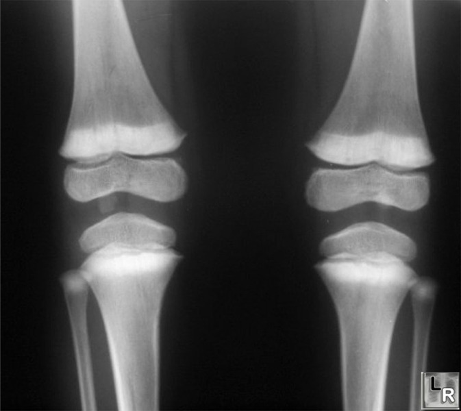 (
Image Source
)
Questions
- Identify the classic radiological finding expected at the metaphyses of long bones and state its underlying histopathologic basis.
- Name the definitive diagnostic test to establish the diagnosis, and state the threshold blood level defining toxicity requiring urgent chelation therapy in children.
- Formulate the combination parenteral chelation regimen indicated for severe lead encephalopathy or blood lead levels $\ge 70\text{ mcg/dL}$.
- State two key cellular mechanisms by which this heavy metal causes microcytic anemia and peripheral neuropathy.
Answer
- Radiological Finding & Basis:
- Finding: Dense, transverse radiopaque bands at the metaphyses of rapidly growing long bones ("lead lines"), predominantly seen at the distal femur, proximal tibia, and distal radius.
- Pathological Basis: Defective resorption of calcified cartilage matrix and hypermineralization with excessive calcium and lead salt deposition in the zone of provisional calcification, rather than pure lead deposition.
- Definitive Diagnostic Test & Chelation Cutoff:
- Test: Venous blood lead level (BLL) measured by atomic absorption spectrometry or anodic stripping voltammetry.
- Threshold for Urgent Chelation: Blood lead level $\ge 45\text{ mcg/dL}$ indicates immediate chelation therapy; BLL $\ge 70\text{ mcg/dL}$ (or any child with lead encephalopathy) represents a critical emergency requiring dual parenteral chelation.
- Dual Chelation Regimen for Severe Poisoning / Encephalopathy ($\ge 70\text{ mcg/dL}$):
- First Agent: Dimercaprol (BAL) $75\text{ mg/m}^2/\text{dose}$ (or $4\text{ mg/kg/dose}$) deep intramuscularly every 4 hours.
- Second Agent: Calcium Disodium Edetate ($\text{CaNa}_2\text{EDTA}$) $1000\text{ to }1500\text{ mg/m}^2/\text{day}$ (or $50\text{ mg/kg/day}$) via continuous intravenous infusion (or divided IV doses every 6–12 hours), started 4 hours after the first dose of BAL (once adequate urine output is established) to avoid mobilizing lead into brain tissue.
- Duration: Continued for 5 consecutive days.
- Cellular Mechanisms:
- Microcytic Anemia: Direct inhibition of critical sulfhydryl-containing heme biosynthetic enzymes ($\delta$-aminolevulinic acid dehydratase and ferrochelatase) and inhibition of pyrimidine 5'-nucleotidase leading to erythrocyte membrane fragility and basophilic stippling.
- Neuropathy: Disruption of calcium-dependent neurotransmitter release, breakdown of the blood-brain barrier via endothelial damage, and demyelination with Schwann cell degeneration.
TOX009 - Toddler Toxic Plant Ingestion
Scenario
A 2-year-old girl (body weight $12\text{ kg}$) is brought to the emergency department after ingesting crushed seeds of an ornamental plant (Nerium oleander) from her garden 3 hours ago. On arrival, she is conscious but nauseated and has vomited twice. Physical examination: heart rate $48\text{ beats/min}$, regular; respiratory rate $24\text{ breaths/min}$; blood pressure $84/52\text{ mmHg}$; capillary blood glucose $98\text{ mg/dL}$; and $\text{SpO}_2\ 99\%$ on ambient air. A 12-lead electrocardiogram (ECG) is obtained.

Questions
- Name the primary cardiotoxic glycosides present in this plant and identify the expected electrocardiographic abnormality described in this child.
- Describe the cellular mechanism by which these plant cardiotoxins induce positive inotropy, delayed conduction, and ventricular ectopy.
- Name the specific definitive neutralizing antidote of choice and state its empiric dosing recommendation when the exact ingested dose is unknown.
- Name one commonly used intravenous electrolyte solution that is strictly contraindicated in this emergency and explain why.
Answer
- Toxins & Electrocardiographic Rhythm:
- Active Toxins: Oleandrin, neriine, and folinerin (cardenolide/cardiac glycosides).
- ECG Abnormality: Severe sinus bradycardia with high-degree (or second-degree) atrioventricular (AV) nodal block, along with scooped/coved ST-segment depression ("reverse tick" or "Salvador Dali mustache" sign).
- Cellular Mechanism of Toxicity:
- Inhibition of the myocardial sarcolemmal $\text{Na}^+/\text{K}^+$-ATPase pump, leading to increased intracellular sodium ($\text{Na}^+$).
- This reverses or suppresses the forward action of the electrogenic $\text{Na}^+/\text{Ca}^{2+}$ exchanger, resulting in marked accumulation of cytosolic free calcium ($\text{Ca}^{2+}$).
- Intracellular calcium overload enhances cardiac contractility and triggers delayed afterdepolarizations (DADs), producing automaticity and arrhythmias, while vagotonic effects and AV nodal conduction delay cause profound bradycardia and heart block.
- Definitive Antidote & Empiric Dosing:
- Antidote: Digoxin-specific antibody fragments (Digoxin-Fab fragments / DigiFab).
- Empiric Dosing (Unknown Ingestion): Administer 10 to 20 vials (400 to 800 mg) IV reconstituted over 30 minutes in acute life-threatening instability; for small pediatric ingestions with known stable presentation, an initial empiric dose of 1 to 2 vials (40 to 80 mg) IV can be titrated against rhythm recovery.
- Contraindicated Electrolyte & Reason:
- Intravenous Calcium (Calcium Gluconate / Calcium Chloride): Strictly contraindicated.
- Reason: Rapid intravenous calcium worsens intracellular hypercalcemia in poisoned cardiomyocytes, precipitating refractory ventricular fibrillation, electromechanical dissociation, and irreversible myocardial contracture ("stone heart").
TOX010 - Adolescent Acute Cerebellar Ataxia
Scenario
A 16-year-old girl is brought to the pediatric emergency department with a 24-hour history of rapidly worsening gait ataxia, incoordination, and slurred speech. Her mother noticed that she had difficulty buttoning her shirt and her handwriting had become illegible. There is no prior history of trauma, fever, or illicit drug abuse. Her father has focal epilepsy treated with daily oral medications kept in the household medicine chest. On examination, the patient is alert but dysarthric, with prominent bilateral horizontal gaze-evoked nystagmus, finger-nose dysmetria, dysdiadochokinesia, and a broad-based ataxic gait.
Questions
- State the most likely diagnosis based on the clinical presentation and household exposure.
- Explain the pharmacokinetic elimination property of this drug that predisposes patients to precipitous toxicity even with minor dose elevations or overdoses.
- List three critical differential diagnoses for acute cerebellar ataxia presenting in this age group that must be excluded.
- Outline the definitive management protocol, including the role of specific antidotes and extracorporeal removal techniques.
Answer
- Diagnosis:
- Acute Phenytoin toxicity (intoxication).
- Pharmacokinetic Elimination Property:
- Michaelis-Menten (Zero-Order / Dose-Dependent / Non-Linear) Kinetics:
- At therapeutic concentrations ($10\text{ to }20\text{ mcg/mL}$), the hepatic cytochrome P450 enzymes (primarily CYP2C9 and CYP2C19) responsible for metabolizing phenytoin become saturated.
- Once saturated, elimination switches from first-order (fixed fraction per unit time) to zero-order (fixed amount per unit time), meaning small increments in dose or ingestion lead to disproportionately large, exponential increases in serum concentrations and prolonged elimination half-life.
- Differential Diagnoses:
- Post-infectious acute cerebellar cerebellitis.
- Posterior fossa lesion (e.g., medulloblastoma, pilocytic astrocytoma, cerebellar abscess, or cerebellar hemorrhage/stroke).
- Alternative acute intoxications (e.g., ethanol, carbamazepine, benzodiazepines, barbiturates, lithium).
- Management Protocol:
- Specific Antidote: None exists; treatment is primarily supportive.
- Supportive Care: Airway, breathing, and circulation maintenance; fall and injury precautions.
- Gastrointestinal Decontamination: Single-dose activated charcoal ($1\text{ g/kg}$, maximum $50\text{ g}$) if presenting within 1–2 hours of acute oral overdose and airway is intact. Multi-dose activated charcoal (MDAC) may accelerate non-renal clearance by interrupting enterohepatic recirculation.
- Extracorporeal Removal: Phenytoin is highly protein-bound ($>90\%$), rendering conventional hemodialysis ineffective. High-flux hemodialysis or hemoperfusion is reserved exclusively for refractory toxicity with hemodynamic collapse or extreme hyperphosphatemia/coma unresponsive to standard intensive supportive care.
More Details
- Phenytoin Concentration-Toxicity Correlation:
- $>20\text{ mcg/mL}$: Far-lateral nystagmus
- $>30\text{ mcg/mL}$: Biphasic nystagmus, marked ataxia, dysmetria, dysarthria
- $>40\text{ mcg/mL}$: Extreme lethargy, stupor, confusion
- $>50\text{ mcg/mL}$: Coma, seizures, paradoxical exacerbation of epileptiform activity
- Note: Intravenous phenytoin formulated in propylene glycol causes hypotension and arrhythmias; oral overdoses cause primarily neuro-vestibular and cerebellar symptoms without direct cardiotoxicity unless massive.
TOX011 - Toddler With Tachypnea And Seizures
Scenario
A 3-year-old boy weighing 14 kg is brought to the pediatric emergency department with a 6-hour history of recurrent non-bilious vomiting, profuse diaphoresis, and altered sensorium culminating in a generalized tonic-clonic seizure lasting 4 minutes. The parents report that he had been staying with his grandparents, where an open, empty bottle of his grandfather's enteric-coated cardiac tablets was discovered. On examination, the child is lethargic and irritable. Vitals: heart rate 148 beats/min, respiratory rate 52 breaths/min (deep, rapid, sighing respirations without chest retractions), blood pressure 92/58 mmHg, axillary temperature 38.6°C, and SpO2 98% on room air.
Arterial Blood Gas (ABG) on room air reveals:
- pH: 7.32
- $\text{pCO}_2$: 20 mmHg
- $\text{pO}_2$: 96 mmHg
- $\text{HCO}_3^-$: 10 mEq/L
- Base Deficit: -14 mEq/L
Serum Biochemistry:
- Sodium ($\text{Na}^+$): 138 mEq/L
- Potassium ($\text{K}^+$): 3.6 mEq/L
- Chloride ($\text{Cl}^-$): 102 mEq/L
- Blood Glucose: 198 mg/dL
Questions
- Interpret the primary acid-base abnormality and explain the dual pathophysiological mechanisms responsible for this mixed disorder.
- Explain the mechanisms responsible for altered glucose homeostasis in this acute ingestion.
- Calculate the Serum Anion Gap and the Delta Ratio ($\Delta \text{AG} / \Delta \text{HCO}_3^-$) to prove the coexisting underlying metabolic derangements.
- Outline the urinary alkalinization protocol, specifying the fluid composition, target urinary and systemic pH, and the mandatory co-infusion requirement.
- Enlist 4 absolute indications for emergent hemodialysis in this patient.
Answer
- Acid-Base Interpretation & Dual Pathophysiology:
- Interpretation: Mixed High Anion Gap Metabolic Acidosis (HAGMA) with primary Respiratory Alkalosis.
- Respiratory Alkalosis Mechanism: Salicylates directly stimulate the medullary respiratory center in the brainstem, causing marked tachypnea, hyperpnea, and excessive pulmonary excretion of $\text{CO}_2$ ($\text{pCO}_2 < 35 \text{ mmHg}$).
- Metabolic Acidosis Mechanism: Salicylates uncouple mitochondrial oxidative phosphorylation, leading to failure of ATP generation, acceleration of glycolysis, accumulation of organic lactic acid and pyruvic acid, mobilization of free fatty acids with accumulation of acetoacetic acid and $\beta$-hydroxybutyrate, and the accumulation of exogenous acidic salicylate metabolites.
- Glucose Homeostasis Dysregulation:
- Hyperglycemia (Early): Caused by chemical stimulation of the adrenal medulla triggering epinephrine release, which accelerates hepatic glycogenolysis and hepatic gluconeogenesis while decreasing peripheral glucose uptake.
- Neuroglycopenia / Hypoglycemia (Late): Mitochondrial uncoupling induces massive cellular glycolysis and depletion of hepatic glycogen stores. Salicylates also impair cellular active glucose transport across the blood-brain barrier, resulting in profound cerebral neuroglycopenia despite normal or elevated peripheral blood glucose levels.
- Mathematical Calculation: $$\begin{aligned} \text{Serum Anion Gap (AG)} &= \text{Na}^+ - (\text{Cl}^- + \text{HCO}_3^-) \\ &= 138 - (102 + 10) \\ &= \mathbf{26\text{ mEq/L}} \quad (\text{Reference: } 8-12\text{ mEq/L}) \end{aligned} $$ $$\begin{aligned} \Delta \text{Ratio} &= \frac{\Delta \text{AG}}{\Delta \text{HCO}_3^-} = \frac{\text{Observed AG} - 12}{24 - \text{Observed HCO}_3^-} \\ &= \frac{26 - 12}{24 - 10} = \frac{14}{14} = \mathbf{1.0} \quad (\text{Reference: } 1.0-2.0 = \text{Pure HAGMA}) \end{aligned} $$
- Urinary Alkalinization Protocol:
- IV Fluid Composition: $5\%$ Dextrose in Water ($\text{D}_5\text{W}$) with $100-150\text{ mEq}$ of Sodium Bicarbonate ($8.4\%$) plus $20-40\text{ mEq}$ of Potassium Chloride ($\text{KCl}$) per liter of infusate.
- Infusion Rate: Administer at $1.5 \text{ to } 2 \times$ maintenance rate ($3-4\text{ mL/kg/hour}$).
- Target Parameters: Urine $\text{pH } \mathbf{7.5 - 8.0}$; maintain arterial systemic blood $\text{pH } \le \mathbf{7.55}$ (stop or titrate down if systemic $\text{pH} > 7.55$).
- Mandatory Potassium Co-infusion: Hypokalemia prevents urinary alkalinization because renal distal tubular $\text{H}^+/\text{K}^+$ ATPase pumps preferentially reabsorb $\text{K}^+$ in exchange for secreting $\text{H}^+$ into urine, resulting in paradoxical aciduria.
- Indications for Emergent Hemodialysis:
- Serum salicylate concentration $> 100\text{ mg/dL}$ ($7.2\text{ mmol/L}$) in acute ingestion (or $> 50-60\text{ mg/dL}$ in chronic toxicity).
- Central nervous system dysfunction (coma, cerebral edema, intractable seizures).
- Acute pulmonary edema / Non-cardiogenic ARDS.
- Refractory acidemia ($\text{pH} < 7.20$) or electrolyte derangements unresponsive to alkalinization.
- Acute renal failure or oliguria preventing therapeutic renal excretion.
More Details
graph TD
    A[Salicylate Ingestion] --> B[Direct Medullary Stimulation]
    A --> C[Uncoupling of Oxidative Phosphorylation]
    B --> D[Hyperpnea / Tachypnea]
    D --> E[Primary Respiratory Alkalosis]
    C --> F[Anaerobic Metabolism & Krebs Inhibition]
    F --> G[Lactic Acid, Pyruvate, Ketoacids]
    G --> H[High Anion Gap Metabolic Acidosis]
    E --> I[Mixed Acid-Base Disorder: pH near normal or acidemic]
    H --> I
    I --> J[Systemic Acidemia Drives Non-ionized Salicylic Acid into CNS]
    J --> K[Cerebral Edema, Seizures, Coma]
    K --> L[Urgent Hemodialysis & NaHCO3 Alkalinization]
TOX012 - Bite Injury With Respiratory Failure
Scenario
A 7-year-old boy weighing 22 kg is brought to the rural emergency department at 04:00 AM from a farm cottage. The parents state that while sleeping on an earthen floor, the child woke up screaming with severe pain in his left foot. Within 3 hours, he developed difficulty opening his eyes, drooling of saliva, and voice change. On examination, the child is anxious and diaphoretic. Vitals: heart rate 104 beats/min, respiratory rate 32 breaths/min with shallow abdominal breathing and intercostal indrawing, blood pressure 106/68 mmHg, and SpO2 86% on room air.
Examination of the left ankle reveals two distinct puncture marks with mild surrounding erythema, minimal edema, and no necrosis or active bleeding. Neurological evaluation reveals bilateral complete ptosis, external ophthalmoplegia, broken single-breath count ($< 7$), pooling of pharyngeal secretions, and inability to lift his head off the bed. A 20-minute whole blood clotting test (20WBCT) demonstrates a firm, solid clot at 20 minutes.
Questions
- Outline the Government of India (GoI) National Snakebite Management Protocol first-aid guideline ("Do It R.I.G.H.T.") and enumerate 4 traditional practices that are strictly contraindicated.
- Outline the immediate airway and ventilatory resuscitation plan for this child.
- Detail the polyvalent Anti-Snake Venom (ASV) administration protocol according to the National Snakebite Management Protocol of India (initial dose, preparation, infusion rate, and criteria for repetition).
- Describe the Neostigmine trial protocol (drugs, dosing, evaluation schedule, and therapeutic continuation criteria).
Answer
- GoI "Do It R.I.G.H.T." First-Aid Guidelines & Contraindications:
- The "Do It R.I.G.H.T." Protocol (GoI / WHO Recommended First Aid):
- R – Reassure the patient: Explain that $\approx 70\%$ of all snakebites are by non-venomous snakes and up to $50\%$ of venomous bites are "dry bites" without envenomation. Keeping the child calm reduces anxiety and prevents tachycardia that hastens systemic venom dissemination.
- I – Immobilize the bitten limb: Splint the affected limb exactly as for a bone fracture using a rigid splint or cloth sling. Avoid walking, running, or any muscular movement of the limb. Keep the limb at or slightly below the level of the heart.
- G.H. – Get to Hospital immediately: Rapidly and smoothly transport the child to a formal medical facility equipped with Polyvalent Anti-Snake Venom (ASV), airway equipment, and mechanical ventilation. Do not waste precious time consulting traditional healers or faith healers.
- T – Tell the doctor: Give a clear, chronological account of symptoms as they emerged (e.g., ptosis, diplopia, dysphagia, voice change, abdominal pain, bleeding) and the exact time of the bite.
- Strictly Contraindicated Traditional Practices:
- Application of tight arterial tourniquets or tight circular cords/ligatures (causes severe ischemia, peripheral nerve damage, gangrene, and massive explosive venom release on sudden removal).
- Making incisions, razor cuts, or scarification over fang marks.
- Oral suctioning or application of vacuum/mechanical suction devices.
- Cryotherapy (ice pack application), cauterization, electric shock therapy, potassium permanganate, or herbal/cow-dung pastes.
- The "Do It R.I.G.H.T." Protocol (GoI / WHO Recommended First Aid):
- Immediate Airway & Ventilatory Resuscitation:
- Airway Clearance: Immediate gentle pharyngeal suctioning to clear pooled secretions; positioning in lateral tilt/recovery position or head-up tilt.
- Oxygenation: High-flow supplemental oxygen via non-rebreather mask ($10\text{--}15\text{ L/min}$).
- Definitive Airway Control: Immediate Rapid Sequence Intubation (RSI) using a cuffed endotracheal tube due to bulbar weakness, loss of airway protective reflexes, and impending diaphragmatic respiratory failure (paradoxical breathing).
- Ventilation: Initiate mechanical ventilation with lung-protective settings ($\text{Vt } 6\text{--}8\text{ mL/kg}$, PEEP $5\text{ cmH}_2\text{O}$) until neuromuscular transmission recovers.
- National Snakebite Protocol - ASV Administration:
- Initial Dose: $\mathbf{10\text{ vials}}$ of lyophilized polyvalent ASV (neutralizes Naja naja, Bungarus caeruleus, Daboia russelii, Echis carinatus).
- Dilution & Preparation: Reconstitute each vial with $10\text{ mL}$ of sterile water. Dilute the 10 vials ($100\text{ mL}$) in $5\text{--}10\text{ mL/kg}$ (maximum $100\text{--}200\text{ mL}$) of Normal Saline ($0.9\% \text{ NaCl}$) or $5\%$ Dextrose.
- Infusion Rate: Infuse slowly at $1\text{ mL/min}$ for the first 10–15 minutes while monitoring for anaphylactoid reactions; if no adverse reaction occurs, complete the remaining infusion over $\mathbf{1\text{ hour}}$.
- Repeat Dose Criteria: In neurotoxic envenomation (Bungarus / Naja), if no objective neurological improvement occurs after $\mathbf{1\text{--}2\text{ hours}}$, administer a second dose of $\mathbf{10\text{ vials}}$ ASV. (Total maximum recommended ASV dose is $20\text{ vials}$; neurotoxic recovery depends primarily on assisted mechanical ventilation as presynaptic $\beta$-neurotoxins cause irreversible terminal axonal damage).
- Neostigmine Trial Protocol:
- Pre-treatment Antimuscarinic: Administer Atropine $0.05\text{ mg/kg}$ IV (or Glycopyrrolate $0.01\text{ mg/kg}$ IV) immediately prior to anticholinesterase administration to prevent cholinergic muscarinic side effects (bradycardia, severe bronchorrhea).
- Neostigmine Dose: Administer Neostigmine $0.04\text{ mg/kg}$ IV (or $0.05\text{ mg/kg}$ IM).
- Evaluation Schedule: Objectively measure neurological indicators (degree of ptosis, single breath count, intercostal muscle movement, neck flexion strength) every $10\text{ to }15\text{ minutes}$ for $\mathbf{30\text{--}60\text{ minutes}}$.
- Interpretation & Maintenance: A positive test is defined by marked improvement in ptosis or respiratory parameters within 30 minutes (more pronounced in postsynaptic Naja bites than presynaptic Bungarus bites). If positive, maintain with Neostigmine $0.01\text{--}0.04\text{ mg/kg}$ IV/IM every $2\text{--}4\text{ hours}$ co-administered with Atropine until complete motor recovery.
More Details
graph TD
    A["Suspected Snakebite (e.g. Bungarus caeruleus)"] --> B["First Aid: Do It R.I.G.H.T."]
    B --> C["R: Reassure | I: Immobilize | GH: Get to Hospital | T: Tell Doctor"]
    C --> D["Assess Signs of Neurotoxicity (Ptosis, Bulbar Palsy, Low Breath Count)"]
    D --> E["Secure Airway: Intubate & Mechanical Ventilation"]
    E --> F["Administer 10 Vials Polyvalent ASV over 1 Hour"]
    F --> G["Neostigmine + Atropine Challenge Test"]
TOX013 - Child With Discrepant Oxygenation Parameters
Scenario
A 5-year-old child presents to the emergency room in acute distress with altered sensorium and perioral/acral cyanosis. Standard transmission pulse oximetry and concurrent arterial blood gas (ABG) co-oximetry yield divergent diagnostic pictures across toxicological exposures. The pediatric intensivist is evaluating three distinct toxicological presentations characterized by mismatched oxygenation profiles.
Questions
- Match each clinical-laboratory oxygenation profile in Column A with the most likely offending toxicant in Column B:
| Column A (Oxygenation Profile) | Column B (Toxic Agents) |
|---|---|
| Profile 1: Marked cyanosis refractory to $100\%$ $\text{O}_2$, pulse oximeter $\text{SpO}_2$ fixed at $85\%$, normal calculated $\text{SaO}_2 \approx 98\%$, normal arterial $\text{PaO}_2 > 90\text{ mmHg}$. | A. Hydrocarbon (kerosene) aspiration pneumonitis |
| Profile 2: Severe lactic acidosis, bright red retinal veins, pulse oximeter $\text{SpO}_2 99\%$, normal arterial $\text{PaO}_2$, markedly elevated $\text{ScvO}_2 > 90\%$. | B. Cyanide ingestion |
| Profile 3: Tachypnea, bilateral crackles, coughing, $\text{SpO}_2 78\%$, true fractional $\text{SaO}_2 77\%$, arterial $\text{PaO}_2 48\text{ mmHg}$ on room air following chemical ingestion/aspiration. | C. Methemoglobinemia-inducing agent (e.g., dapsone, benzocaine, aniline dyes) |
- Explain the physical principle why standard dual-wavelength transmission pulse oximeters display an $\text{SpO}_2$ fixed around $85\%$ in Profile 1.
- What is the classic bedside visual test on blood drawn in Profile 1, and what is the definitive antidote and its specific contraindication?
- Explain the cellular mechanism of lactic acidosis and high $\text{ScvO}_2$ in Profile 2, and name two specific antidotal therapies.
Answer
- Matching Table:
| Profile (Column A) | Offending Toxicant (Column B) | Pathophysiological Hallmark |
|---|---|---|
| Profile 1 | C. Methemoglobinemia-inducing agent | Dual-wavelength pulse oximeter saturates at $\approx 85\%$ due to $1:1$ absorbance ratio at $660/940\text{ nm}$; normal dissolved plasma $\text{PaO}_2$. |
| Profile 2 | B. Cyanide ingestion | Histotoxic cellular hypoxia via mitochondrial cytochrome c oxidase (Complex IV) inhibition; tissues cannot extract $\text{O}_2$, leading to high $\text{ScvO}_2$. |
| Profile 3 | A. Hydrocarbon pneumonitis | Alveolar epithelial necrosis, surfactant disruption, and severe ventilation-perfusion ($V/Q$) mismatch causing true hypoxemia. |
- Physical Basis of Pulse Oximeter Reading in Methemoglobinemia:
- Standard dual-wavelength pulse oximeters measure light absorption at two specific wavelengths: $660\text{ nm}$ (red) and $940\text{ nm}$ (infrared).
- Methemoglobin has an extraordinarily high absorption coefficient at both $660\text{ nm}$ and $940\text{ nm}$.
- When methemoglobin levels exceed $30\text{ to }35\%$, the absorption ratio ($R = A_{660} / A_{940}$) approaches $1.0$.
- In standard calibration algorithms, an absorption ratio of $1.0$ corresponds mathematically to an oxygen saturation of $85\%$. Thus, regardless of true arterial oxyhemoglobin, the pulse oximeter reading plateaus around $85\%$.
- In contrast, routine ABG machines calculate saturation indirectly from $\text{PaO}_2$ (which is dissolved plasma oxygen and remains normal), producing a falsely normal calculated saturation. Definitive diagnosis requires co-oximetry.
- Bedside Test, Antidote, and Contraindication (Profile 1):
- Bedside Visual Test: Place a drop of the patient blood on filter paper (or white gauze) next to normal control blood. Methemoglobinemic blood appears chocolate brown / slate-gray and does NOT turn bright red upon blowing $100\%$ oxygen over it (unlike deoxygenated normal hemoglobin).
- Definitive Antidote: Methylene Blue ($1\%$ solution; $1\text{ to }2\text{ mg/kg}$ IV infused slowly over 5 minutes; acts as an electron donor for NADPH-dependent methemoglobin reductase).
- Contraindication: Glucose-6-Phosphate Dehydrogenase (G6PD) deficiency (Methylene blue requires NADPH generated by the pentose phosphate pathway; in G6PD deficiency, it is ineffective and can induce massive acute intravascular hemolysis). Alternative therapy: High-dose Ascorbic acid (Vitamin C) or Exchange Transfusion.
- Mechanism and Antidotes for Cyanide (Profile 2):
- Cellular Mechanism:
- Cyanide binds with high affinity to the ferric ($\text{Fe}^{3+}$) iron of mitochondrial cytochrome c oxidase (Complex IV) in the electron transport chain.
- This halts aerobic oxidative phosphorylation, preventing cellular utilization of oxygen despite normal arterial delivery and normal hemoglobin saturation.
- Cells shift to anaerobic glycolysis, generating massive amounts of lactic acid (severe lactic acidosis). Unextracted oxygen remains in venous blood, resulting in arterialization of venous blood and high central venous saturation ($\text{ScvO}_2 > 90\%$).
- Antidotal Therapies:
- Hydroxocobalamin (Cyanokit): First-line antidote ($70\text{ mg/kg}$ IV over 15 minutes; binds cyanide directly to form nontoxic cyanocobalamin, excreted in urine).
- Sodium Nitrite + Sodium Thiosulfate: Sodium nitrite ($10\text{ mg/kg}$ IV) induces controlled methemoglobinemia ($\text{Fe}^{3+}$), which scavenges cyanide from cytochrome oxidase to form cyanomethemoglobin; Sodium thiosulfate ($1.65\text{ mL/kg}$ of $25\%$ solution IV) acts as a sulfur donor for rhodanese to convert cyanide into thiocyanate.
- Cellular Mechanism:
TOX014 - Pediatric Toxidromes, Antidotes, And Toxicokinetics
Scenario
A 4-year-old child is rushed to the pediatric emergency triage with acute altered sensorium, erratic vital signs, and suspected unwitnessed ingestion of household chemicals or grandparental medications 2 hours prior to arrival. The pediatric emergency team is simultaneously performing rapid toxidrome recognition, selecting targeted antidotes, assessing toxicokinetics through osmolality calculations, and considering gastrointestinal decontamination.
Questions
- Match each clinical toxidromic presentation in Column A with the most likely offending toxicant or drug class in Column B:
| Column A (Toxidromic Presentation) | Column B (Offending Toxicants) |
|---|---|
| Presentation A: Pinpoint pupils, bradycardia, diaphoresis, profuse bronchorrhea, muscle fasciculations, urinary incontinence. | I. Isoniazid (INH) |
| Presentation B: Hyperpyrexia, non-reactive mydriasis, flushed/dry skin, absent bowel sounds, urinary retention, purposeless picking movements. | II. Salicylates (Aspirin) |
| Presentation C: High anion gap metabolic acidosis, hyperglycemia, status epilepticus refractory to standard first-line benzodiazepines. | III. Organophosphates / Carbamates |
| Presentation D: Hyperpnea (central hyperventilation), high anion gap metabolic acidosis, tinnitus, hyperthermia, non-cardiogenic pulmonary edema. | IV. Datura stramonium / Anticholinergics |
- Complete the pharmacological antidote profile for the following toxic exposures:
- Beta-blocker overdose
- Isoniazid (INH) acute poisoning
- Acquired toxic methemoglobinemia
- Acute elemental iron toxicity
- The child's stat metabolic workup yields: Sodium ($\text{Na}^+$) = $138\text{ mEq/L}$, Blood Glucose = $90\text{ mg/dL}$, Blood Urea Nitrogen ($\text{BUN}$) = $14\text{ mg/dL}$, and Measured Serum Osmolality = $325\text{ mOsm/kg } \text{H}_2\text{O}$. Calculate the Serum Osmolar Gap and interpret the result.
- Outline the procedural checklist, indications, and absolute contraindications for administering Single-Dose Activated Charcoal (SDAC) in a pediatric emergency setting.
Answer
- Toxidrome-to-Toxin Matching Table:
| Presentation (Column A) | Matched Toxicant (Column B) | Underlying Toxidrome & Mechanism |
|---|---|---|
| Presentation A | III. Organophosphates / Carbamates | Cholinergic Toxidrome (SLUDGEM/DUMBELS): Acetylcholinesterase inhibition producing muscarinic and nicotinic hyperstimulation. |
| Presentation B | IV. Datura / Anticholinergics | Anticholinergic Toxidrome: Muscarinic receptor antagonism ("blind as a bat, mad as a hatter, red as a beet, hot as a hare, dry as a bone"). |
| Presentation C | I. Isoniazid (INH) | GABA Deficiency Toxidrome: Depletion of pyridoxal-5-phosphate (PLP) inhibiting GABA synthesis, causing intractable seizures and severe lactic acidosis. |
| Presentation D | II. Salicylates (Aspirin) | Salicylate Toxidrome: Direct medullary respiratory stimulation plus mitochondrial oxidative phosphorylation uncoupling. |
- Pharmacological Antidote Profile Table:
| Toxic Exposure | Specific Antidote | Mechanism of Action | Pediatric Dosing & Administration |
|---|---|---|---|
| Beta-blocker overdose | Glucagon (companion: HIET) | Bypasses adrenergic receptors; directly stimulates adenylate cyclase via G-protein-coupled glucagon receptors, raising intracellular cAMP. | $50\text{--}150\ \mu\text{g/kg}$ IV bolus over 1–2 min (max $10\text{ mg}$), then infusion at $0.05\text{--}0.1\text{ mg/kg/h}$. Companion: High-Dose Insulin Euglycemia Therapy (HIET). |
| Isoniazid (INH) | Pyridoxine (Vitamin $\text{B}_6$) | Restores pyridoxal-5-phosphate (PLP) cofactor essential for glutamic acid decarboxylase (GAD) to synthesize inhibitory GABA. | Gram-for-gram equivalent to ingested INH dose IV; if unknown, $70\text{ mg/kg}$ IV (max $5\text{ g}$) infused at $0.5\text{ g/min}$. |
| Toxic Methemoglobinemia | Methylene Blue (1% solution) | Exogenous cofactor reduced by NADPH-dependent methemoglobin reductase to leucomethylene blue, which non-enzymatically reduces $\text{Fe}^{3+}$ to $\text{Fe}^{2+}$. | $1\text{--}2\text{ mg/kg}$ ($0.1\text{--}0.2\text{ mL/kg}$ of 1% solution) IV slow push over 5 min. (Contraindicated in G6PD deficiency). |
| Acute Iron Toxicity | Deferoxamine mesylate | Chelates free circulating ferric iron ($\text{Fe}^{3+}$) forming water-soluble, non-toxic ferrioxamine excreted in urine ("vin rosé" color). | Continuous IV infusion starting at $15\text{ mg/kg/h}$ (max $40\text{ mg/kg/h}$ in severe shock; daily limit $6\text{ g}$) until serum iron $<350\ \mu\text{g/dL}$. |
Serum Osmolar Gap Derivation:
$$\begin{aligned} \text{Calculated Serum Osmolality} &= 2 \times [\text{Na}^+] + \frac{[\text{Glucose}]}{18} + \frac{[\text{BUN}]}{2.8} \\ &= 2 \times 138 + \frac{90}{18} + \frac{14}{2.8} \\ &= 276 + 5 + 5 = 286\text{ mOsm/kg } \text{H}_2\text{O} \\ \text{Osmolar Gap} &= \text{Measured Osmolality} - \text{Calculated Osmolality} \\ &= 325 - 286 \\ &= \mathbf{39\text{ mOsm/kg } \text{H}_2\text{O}} \quad (\text{Normal: } < 10\text{ mOsm/kg } \text{H}_2\text{O}) \end{aligned} $$- Clinical Interpretation: Elevated Osmolar Gap ($>10\text{ mOsm/kg}$) signifies the systemic presence of unmeasured, low-molecular-weight, osmotically active exogenous compounds, primarily toxic alcohols (methanol, ethylene glycol, isopropanol) or propylene glycol.
Single-Dose Activated Charcoal (SDAC) Protocol Table:
| Clinical Parameter | Guideline Recommendations |
|---|---|
| Indications & Timing | Ingestion of a potentially lethal, charcoal-adsorbable substance presenting within 1 hour of ingestion. |
| Pediatric Dose & Route | $1\text{ g/kg}$ (or $25\text{--}50\text{ g}$ in older children) administered orally or via large-bore nasogastric tube as an aqueous slurry. |
| Safety Checklist | Confirm intact gag/airway protective reflexes (or secure with cuffed ETT); verify presence of active bowel sounds. |
| Absolute Contraindications | • Unprotected airway / depressed sensorium (severe aspiration pneumonitis risk). • Ingestion of non-adsorbing toxins: PHAILS (Pesticides/Hydrocarbons, Heavy metals [Iron, Lead], Acids/Alkalis/Caustics, Alcohols, Lithium, Solvents). • Gastrointestinal obstruction, perforation, or ileus. |
TOX015 - Toddler With Wide Complex Tachycardia
Scenario
A 14-year-old boy (weight 16 kg) is brought to the pediatric emergency resuscitation bay 90 minutes after an unsupervised ingestion of an unknown quantity of his father's psychiatric prescription medication. On arrival, he alternates between lethargy and delirium. Physical examination reveals warm, flushed, dry skin, bilaterally dilated pupils (6 mm) with sluggish light reflex, absent bowel sounds, and a palpable suprapubic bladder distension.
Initial vitals: Heart rate 132 beats/min, blood pressure 74/40 mmHg, respiratory rate 24 breaths/min, temperature 38.8°C, and SpO₂ 96% on ambient air. A 12-lead electrocardiogram (ECG) is obtained immediately upon admission.
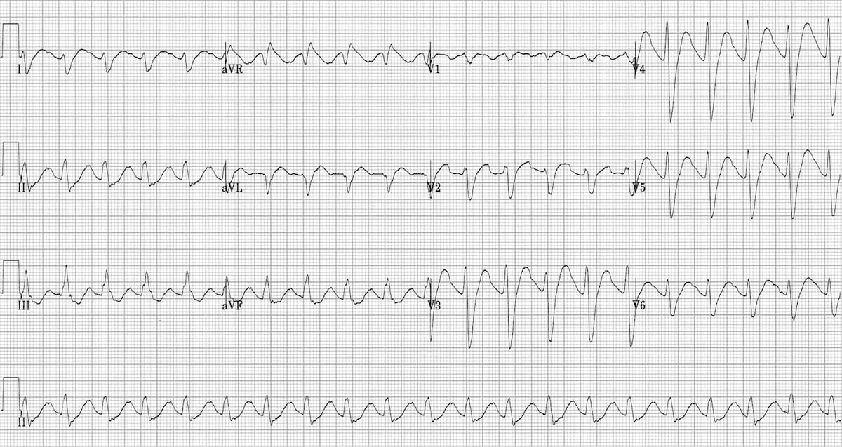 (
Image Source
)
Questions
- Interpret the cardinal findings demonstrated on this 12-lead electrocardiogram.
- Identify the clinical toxidrome and name the offending pharmacological drug class.
- Enlist the classical electrocardiographic criteria used to stratify severity in this poisoning, citing the specific cutoffs for QRS duration and lead aVR morphology that predict adverse outcomes.
- Name two distinct toxicological differentials that mimic the peripheral autonomic features of this toxidrome.
- Outline the definitive first-line emergency pharmacotherapy for the cardiac conduction defects and hypotension, including exact dosing, target therapeutic endpoints, and two pharmacological agents strictly contraindicated in this setting.
Answer
Electrocardiogram Interpretation:
- Wide complex tachycardia with loss of discernible P waves.
- Marked QRS complex prolongation ($>120\text{ ms}$).
- Right axis deviation of the terminal 40 ms frontal QRS vector, manifested as a prominent, wide terminal R wave in lead aVR ($R\text{ wave} \ge 3\text{ mm}$ or $R/S\text{ ratio} > 0.7$).
- Deep, slurred S waves in leads I and aVL; secondary ST-T wave discordance and prolonged QTc interval.
Clinical Toxidrome & Drug Class:
- Toxidrome: Anticholinergic toxidrome combined with cardiac fast sodium-channel blockade (quinidine-like cardiotoxicity).
- Drug Class: Tricyclic Antidepressants (TCAs; e.g., amitriptyline, imipramine, nortriptyline).
ECG Risk Stratification Criteria:
- QRS Interval Duration:
- $\text{QRS} > 100\text{ ms}$: Predicts an approximate 33% risk of generalized seizures.
- $\text{QRS} > 160\text{ ms}$: Predicts an approximate 50% risk of life-threatening ventricular dysrhythmias (monomorphic VT, polymorphic VT, ventricular fibrillation).
- Terminal 40-ms QRS Axis in Lead aVR:
- Terminal R wave amplitude $\ge 3\text{ mm}$ in aVR.
- $R/S\text{ ratio}$ in lead aVR $> 0.7$ (strongly correlates with impending ventricular dysrhythmias and shock).
- Conduction Delays: Prolonged PR interval, non-specific intraventricular conduction delay (IVCD), and prominent QTc prolongation.
- QRS Interval Duration:
Toxicological Differential Diagnoses:
- Datura stramonium / Belladonna alkaloids / Anticholinergics (atropine, scopolamine, first-generation antihistamines such as diphenhydramine).
- Sympathomimetic agents (Cocaine, amphetamines, ephedrine).
Emergency Pharmacotherapy Protocol:
- First-line Agent: Hypertonic Sodium Bicarbonate ($8.4\%$ solution; provides $1\text{ mEq/mL}$ of $\text{Na}^+$ and $\text{HCO}_3^-$).
- Initial Intravenous Bolus: $$\begin{aligned} \text{Dose} &= 1 \text{ to } 2 \text{ mEq/kg IV push over } 2\text{ to } 3 \text{ minutes} \\ &= 1 \text{ to } 2 \text{ mL/kg of } 8.4\% \text{ solution} \\ &= \mathbf{16 \text{ to } 32 \text{ mL}} \quad (\text{for a } 16\text{-kg child}) \end{aligned} $$ Repeat every 3–5 minutes until QRS complex narrows ($<100\text{ ms}$) and systolic blood pressure normalizes.
- Continuous Infusion: $150\text{ mEq}$ of $\text{NaHCO}_3$ in $1\text{ L}$ of $5\%$ Dextrose infused at $1.5\text{ to }2 \text{ times}$ maintenance rate ($2\text{ to }3\text{ mL/kg/h}$).
- Target Physiological Endpoints:
- Arterial blood gas target $\text{pH}: \mathbf{7.45 - 7.55}$ (do not exceed $\text{pH } 7.55$).
- Serum bicarbonate: $30 - 32\text{ mEq/L}$.
- Serum potassium maintained at $4.0 - 4.5\text{ mEq/L}$ (alkalemia induces intracellular potassium shifting).
- Strictly Contraindicated Agents:
- Class IA and IC antiarrhythmics (procainamide, quinidine, flecainide): Worsen sodium channel blockade.
- Class III antiarrhythmics (amiodarone, sotalol): Prolong repolarization and trigger torsades de pointes.
- Physostigmine: Absolutely contraindicated in TCA overdose; precipitates refractory asystole, complete heart block, and intractable seizures.
More Details
Pathophysiologic Mechanisms of Tricyclic Antidepressant (TCA) Overdose
TCAs exhibit a multifaceted toxicological profile mediated via five major pharmacological actions:
flowchart TD
    Top["Tricyclic Antidepressant (TCA) Toxicity Multi-Target Mechanisms"] --> M1["Myocardial Fast Na⁺ Channels Inhibition"]
    Top --> M2["Anticholinergic Blockade (M1)"]
    Top --> M3["Adrenergic Alpha-1 Blockade"]
    Top --> M4["Inhibition of NE / 5-HT Reuptake"]
    Top --> M5["Central GABA-A Antagonism"]

    M1 --> C1["• QRS prolongation (> 100 ms)<br/>• Terminal R wave in aVR (> 3 mm)<br/>• Ventricular dysrhythmias<br/>• Negative inotropy & hypotension"]
    M2 --> C2["• Hyperthermia & anhidrosis<br/>• Mydriasis & blurred vision<br/>• Dry, flushed skin<br/>• Paralytic ileus & urinary retention"]
    M3 --> C3["• Peripheral vasodilation<br/>• Orthostatic hypotension<br/>• Refractory vasodilatory shock"]
    M4 --> C4["• Initial transient hypertension<br/>• Sinus tachycardia<br/>• Arrhythmogenesis"]
    M5 --> C5["• Delirium & agitation<br/>• Myoclonus & hyperreflexia<br/>• Status epilepticus"]
Mechanism of Sodium Bicarbonate Reversal
- Sodium Ion Loading: Overcomes the competitive, voltage-dependent blockade of cardiac fast inward $\text{Na}^+$ channels ($\text{Nav}1.5$) by increasing the extracellular-to-intracellular $\text{Na}^+$ electrochemical gradient.
- Systemic Alkalization: Decreases the active, protonated (ionized) fraction of the lipophilic TCA drug molecule, promoting dissociation of the drug from its receptor site on the sodium channel.
Refractory Hemodynamic Instability Algorithm
- Hypotension refractory to fluid boluses ($10-20\text{ mL/kg}$ balanced crystalloid) and $\text{NaHCO}_3$:
- Initiate direct-acting alpha-adrenergic vasopressors: Norepinephrine (preferred; $0.05-1\text{ mcg/kg/min}$) or Epinephrine. Dopamine is ineffective due to endogenous myocardial catecholamine depletion.
- Intravenous Lipid Emulsion (ILE) 20%: Acts as a "lipid sink" for lipophilic TCAs:
- Initial bolus: $1.5\text{ mL/kg}$ over $2-3\text{ minutes}$.
- Continuous infusion: $0.25\text{ mL/kg/min}$ for $30-60\text{ minutes}$.
- Seizures: Treat aggressively with intravenous Benzodiazepines (Midazolam $0.1-0.2\text{ mg/kg}$ or Lorazepam $0.1\text{ mg/kg}$). Avoid Phenytoin (a class IB antiarrhythmic that worsens cardiac conduction delays).